Importance and Purpose of Microscopic Urine Examination
Microscopic urine examination is crucial for diagnosing urinary tract infections (UTIs) and renal diseases. It detects cells, casts, and crystals, aiding in early detection and management of genitourinary disorders, guiding treatment and monitoring disease progression effectively.
1.1 Role in Diagnosing Urinary Tract Infections (UTIs)
Microscopic urine examination plays a pivotal role in diagnosing UTIs by identifying key components such as leukocytes, red blood cells, and bacteria. The presence of these elements helps confirm infections and assess severity. Timely analysis under a microscope ensures accurate detection of pathogens, guiding appropriate antibiotic treatment. This method is complemented by qualitative urine culture, enhancing diagnostic accuracy. Regular examination also aids in monitoring infection resolution and preventing complications. Proper specimen collection and centrifugation are critical for reliable results, making microscopic analysis indispensable in UTI management and patient care.
1.2 Significance in Renal and Genitourinary Disease Management
Microscopic urine examination is vital for managing renal and genitourinary diseases. It detects abnormalities like casts, crystals, and proteinuria, indicating kidney dysfunction or damage. This method aids in identifying conditions such as glomerulonephritis, pyelonephritis, and nephrotic syndrome, enabling early intervention. By analyzing urine sediment, healthcare providers can monitor disease progression and adjust treatments accordingly. Microscopic findings also guide the diagnosis of asymptomatic conditions, such as microscopic hematuria, which may signal underlying renal or urinary tract issues. Regular examination ensures timely detection of complications, improving patient outcomes and facilitating personalized care plans.
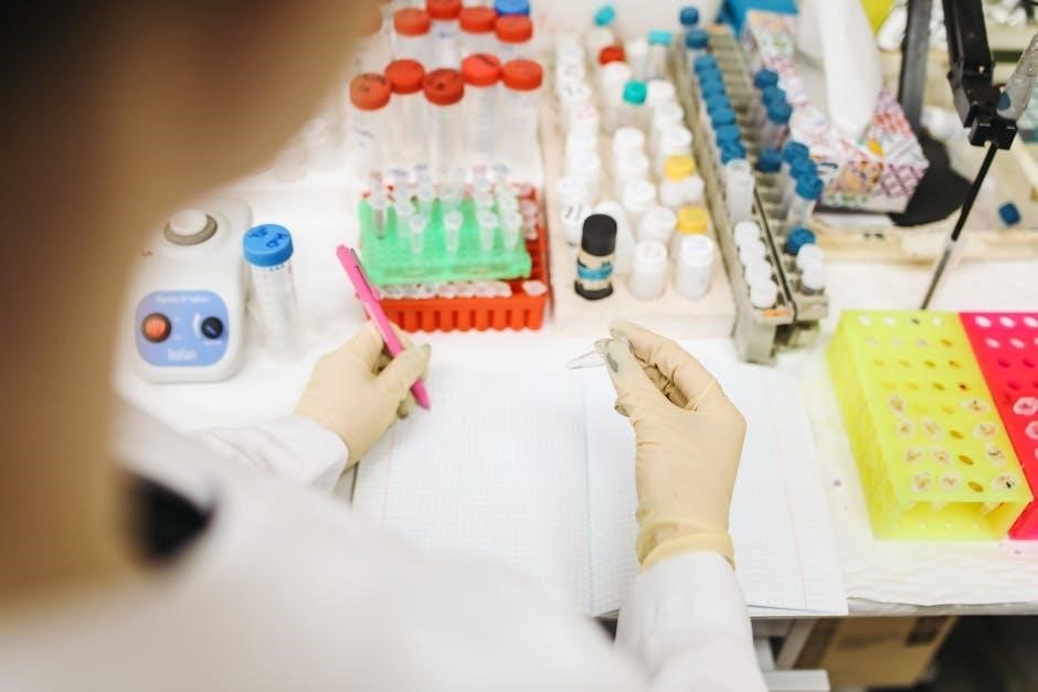
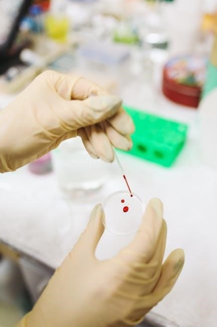
Specimen Collection and Preparation
Proper collection and preparation of urine samples are essential for accurate microscopic examination. This involves using 10-15 ml of well-mixed urine, centrifugation to concentrate sediment, and ensuring freshness to prevent contamination. Proper handling and techniques are crucial to avoid errors and obtain reliable results.

2.1 Proper Patient Preparation and Instructions
Proper patient preparation is vital for accurate microscopic examination. Patients should be instructed to avoid strenuous exercise 48 hours prior to sample collection. They should also avoid sexual activity, douching, or using vaginal creams to prevent contamination. Women should not collect samples during menstruation. Patients should be advised to provide a first-catch morning urine sample, as it is typically more concentrated. Clear instructions should be given to ensure the sample is collected cleanly, using a sterile container if possible. Adherence to these guidelines helps ensure the quality and reliability of the urine sample for microscopic analysis.
2.2 Techniques for Collecting a Clean, Representative Sample

Collecting a clean, representative urine sample is essential for accurate microscopic examination. The midstream urine collection method is recommended to minimize contamination. Patients should start urinating, then collect the middle portion of the stream in a sterile container. The genital area should be cleaned with a sterile wipe before collection to reduce contamination from bacteria, cells, or other debris. For women, the labia should be spread to prevent urine from coming into contact with the vulva. The container should be tightly sealed and labeled promptly. Proper collection ensures the sample reflects the true condition of the urinary tract, avoiding false results.
2.3 Centrifugation Process for Concentrating Urine Sediment
The centrifugation process involves spinning a 10-15 ml urine sample at 1,500-2,000 rpm for 5 minutes to concentrate sediment. This step separates cells, casts, and crystals from the supernatant. After centrifugation, the supernatant is decanted, leaving a concentrated pellet. The sediment is then resuspended in a small amount of residual urine for slide preparation. This method ensures that microscopic examination accurately reflects the urinary tract’s cellular and structural components, enhancing the detection of abnormalities such as infections or kidney disease.
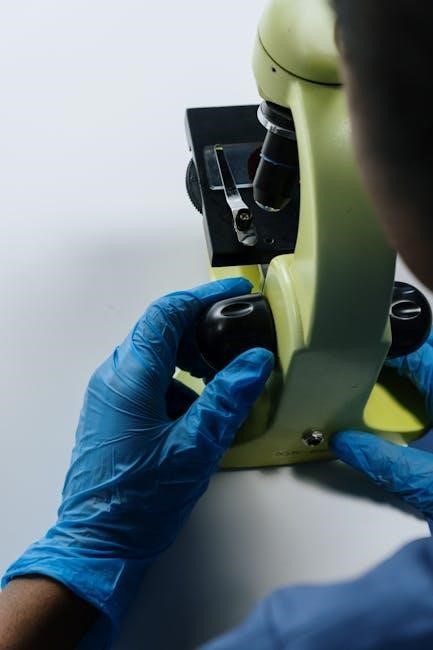
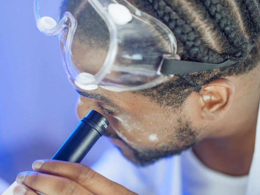
Microscopic Examination Techniques
Microscopic examination involves using a microscope to analyze urine sediment for cells, casts, crystals, and microorganisms. Proper techniques ensure accurate detection of abnormalities in urinary tract health.
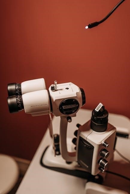
3.1 Slide Preparation Methods
Slide preparation begins with centrifuging 10-15 ml of well-mixed urine at 1,500-2,000 rpm for 5-10 minutes to concentrate sediment. The supernatant is discarded, leaving 0.5-1 ml. A drop of concentrated sediment is placed on a labeled slide, covered with a coverslip. Stains like methylene blue or Sudan III may be applied for better visibility. Proper technique ensures even distribution of particles, avoiding air bubbles. The slide is then ready for microscopic examination under low and high power to identify cells, casts, crystals, and microorganisms. Accurate preparation is critical for reliable diagnostic results.
3.2 Examination Under the Microscope: Key Observations
Under the microscope, urine sediment is examined for cells, casts, crystals, and microorganisms. Red blood cells (RBCs) indicate hematuria, while white blood cells (WBCs) suggest infection. Epithelial cells may be present, but their significance varies. Hyaline casts are normal, whereas granular or red blood cell casts suggest pathology. Crystals like calcium oxalate or uric acid are common but may indicate stone formation. Bacteria, yeast, or parasites confirm infections. Microscopic findings are correlated with clinical symptoms and other tests to aid diagnosis. Accurate observation requires proper focus, illumination, and experience to avoid misinterpretation of artifacts or contaminants.
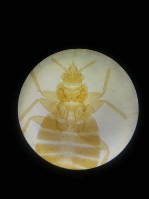
Identification of Urine Sediment Components
Urine sediment components include cells, casts, crystals, and other particles. Cells such as RBCs, WBCs, and epithelial cells are identified, along with casts like hyaline or granular types. Crystals, including calcium oxalate or uric acid, are common. Other structures like bacteria, yeast, or parasites may also be present, aiding in diagnosis.
4.1 Types of Cells and Their Clinical Significance
Microscopic examination identifies various cells in urine, including red blood cells (RBCs), white blood cells (WBCs), and epithelial cells. RBCs indicate hematuria, potentially signaling kidney or urinary tract issues. WBCs suggest infection or inflammation, such as in UTIs. Epithelial cells, often shed from the urinary tract lining, may indicate infection or irritation. The presence and quantity of these cells help diagnose conditions like pyelonephritis or glomerulonephritis. Additionally, abnormal cells, such as cancer cells, may be detected, aiding in early diagnosis of genitourinary malignancies. Other structures like bacteria, yeast, or parasites can also be observed, further guiding clinical decisions and treatment plans.
4.2 Casts: Characteristics and Diagnostic Value
Casts are cylindrical structures formed in renal tubules, composed of proteins and cells. Their presence in urine sediment is diagnostic, often indicating kidney disease. Hyaline casts, the most common type, are usually benign and may appear after physical exertion. Granular or epithelial cell casts suggest tubular damage, while red blood cell casts indicate glomerulonephritis or other glomerular injuries. Casts provide critical insights into renal function and pathology, aiding in the differentiation between prerenal, renal, and postrenal causes of disease. Their characteristics, such as color, consistency, and composition, help guide further diagnostic testing and treatment plans.
4.3 Crystals: Common Types and Their Implications
Crystals in urine can indicate metabolic disorders or kidney stone formation. Common types include calcium oxalate, uric acid, and phosphate crystals. Calcium oxalate crystals are often associated with hypercalciuria and kidney stones, appearing as small, square, or octagonal structures. Uric acid crystals, typically yellow or reddish, are linked to hyperuricemia and gout. Phosphate crystals, seen in alkaline urine, may suggest urinary tract infections or renal tubular damage. The presence and type of crystals provide valuable diagnostic clues, aiding in the assessment of metabolic conditions and guiding preventive measures for stone formation. Accurate identification is essential for targeted clinical management.
4.4 Other Structures and Particles in Urine Sediment
Beyond cells, casts, and crystals, urine sediment may contain other structures like mucus, fungi, parasites, and artificial elements. Mucus threads are normal but increase in UTIs. Fungal elements, such as Candida, indicate infections, especially in immunocompromised patients. Parasites like Trichomonas or Schistosoma are rare but significant. Pollen grains or starch may appear due to contamination. Fibers or lint from collection materials are common artifacts. Identifying these structures helps differentiate between pathological and benign conditions, ensuring accurate diagnoses and appropriate treatment plans. Their presence must be interpreted in the clinical context to avoid misdiagnosis.

Clinical Relevance and Interpretation
Microscopic urine examination aids in diagnosing UTIs, renal diseases, and asymptomatic hematuria, guiding further testing and treatment, ensuring accurate patient care and management of genitourinary conditions.
5.1 Correlating Microscopic Findings with Urinary Tract Infections

Microscopic findings such as increased leukocytes, red blood cells, and bacteria in urine sediment are key indicators of urinary tract infections (UTIs). These components help confirm the presence of infection and guide appropriate antimicrobial therapy. The detection of white blood cells, particularly, signifies the body’s immune response to infection. Additionally, the presence of casts or crystals may further support the diagnosis, aiding clinicians in distinguishing between upper and lower urinary tract infections. Accurate interpretation of these findings ensures timely and targeted treatment, improving patient outcomes and reducing the risk of complications.
5.2 Microscopic Hematuria: Implications and Further Testing
Microscopic hematuria, characterized by the presence of red blood cells in urine, often signals underlying conditions such as kidney stones, infections, or malignancies. While asymptomatic in many cases, it necessitates thorough evaluation to rule out serious pathology. Further testing includes imaging studies like ultrasound or CT scans to visualize the urinary tract. In some cases, cystoscopy or biopsy may be required to investigate the cause. Prompt diagnostic workup ensures early detection and management of potential genitourinary diseases, preventing progression and improving long-term patient outcomes through targeted interventions tailored to the identified cause.
Guidelines and Best Practices
Adherence to European Urinalysis Guidelines ensures accurate microscopic examinations. Proper specimen collection, slide preparation, and avoiding common errors are essential for reliable and consistent diagnostic results.
6.1 European Urinalysis Guidelines for Microscopic Examination
The European Urinalysis Guidelines emphasize standardized methods for microscopic urine examination. They recommend timely analysis of fresh samples, ideally within one hour of collection, to avoid degradation. Proper centrifugation techniques, using 10-15 ml of well-mixed urine at 400-600 rpm for 5-10 minutes, are highlighted. Guidelines also stress the use of preservatives like glutaraldehyde or formaldehyde for delayed analyses. Additionally, they advocate for the use of certified professionals and standardized reporting formats to ensure consistency and accuracy in results. Adherence to these guidelines enhances diagnostic reliability and supports effective clinical decision-making in urinary tract and renal disease management.
6.2 Preservation Techniques for Fresh Urine Samples
Preserving fresh urine samples is critical for accurate microscopic examination. Common techniques include adding preservatives like glutaraldehyde or formaldehyde to prevent cellular degradation. These chemicals stabilize elements, ensuring reliable results even when analysis is delayed. Additionally, storing samples in a cool, dark place can slow bacterial growth and maintain sample integrity. Proper preservation is essential for detecting cells, casts, and crystals, as delays without preservation can lead to inaccurate findings. Timely analysis, ideally within one hour, remains ideal, but preservatives extend the window for precise diagnostic evaluation in clinical and laboratory settings.
6.3 Avoiding Common Errors in Microscopic Analysis
Common errors in microscopic urine analysis include delayed examination, improper centrifugation, and misidentification of sediment components. To avoid these, ensure timely analysis, as cellular changes occur rapidly. Properly centrifuge samples to concentrate sediment effectively. Use standardized techniques for slide preparation to maintain consistency. Train personnel to distinguish between artifacts and pathological elements, such as crystals versus casts. Regularly calibrate microscopes and use reference materials to improve accuracy. Document findings systematically to avoid oversight. Adhering to these practices enhances the reliability of microscopic examination, ensuring accurate diagnosis and treatment in clinical settings.